Lisfranc Fractures and Injuries - Minimally Invasive Surgical Treatment
Lisfranc fractures can occur from a minor misstep, a small twisting injury, or a sports related injury. In some cases, the injury is so subtle that it may be missed on the initial x-rays and the treatment may be delayed.
This injury involves the midfoot, which contains the tarsometatarsal joint. This joints is the where the cuneiform bones and the bases of the metatarsals are joined at the base of the toes. Because of the complex anatomy of this area, Lisfranc injuries can be subtle and difficult to diagnose on plain x-rays. CT scans are usually needed.
| Minimally invasive procedures are now available for lisfranc injuries. If you would like more information on how these treatments may help you with your particular health concerns, please contact our office. |
 |
Case presentation:
In this particular case, a generally healthy, active 66-year-old man missed a step in one of Boston's historic buildings.
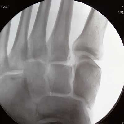
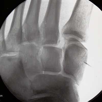
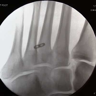
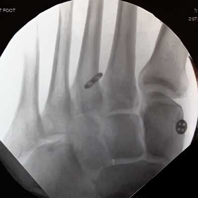
On the initial x-ray, an abnormal space between the bones at the base of the first and second toes can be seen. This is the hallmark of a Lisfranc joint injury or fracture. This xray was taken during the actual surgery, just before the minimally invasive treatment was performed. The patient is comfortable, lying on the operating table under mild sedation and the foot has been blocked with numbing medicine, so that the patient feels no pain. |
. |
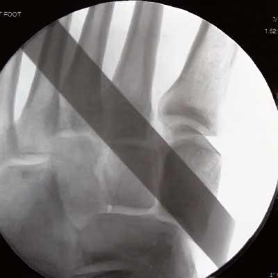
In this next x-ray, a metal ruler is being used to indicate the position of the intended minimally invasive fixation. The top edge of the ruler is exactly where the fixation needs to be. This allows the skin to be marked in the exact location where a tiny incision can be made to place the suture button fixation that is planned for this surgery. Experience is necessary to insure the correct location. |
|
| |
After the skin is marked, a numbing needle is used to identify the exact location on the bone where a guidepin should be placed. The incision is made exactly at that location. The incision is 1/4 long, approximately a height of two quarters stacked on top of each other. |
|
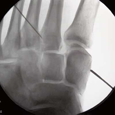
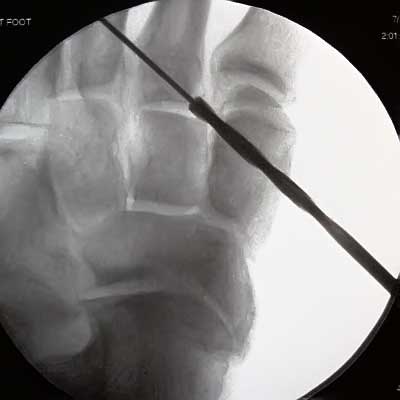
A second small needle is used to identify the location where the guide pin should pass out of the bone. This allows precision targeting of the pin. In this image, The second guided needle is on the top left, and the guide pin is being placed from right to left. |
In the image above, the guide pin has passed through the cuneiform bone and is in the space between the cuneiform and the second metatarsal base. This space is not normally so large. The abnormal spreading of these bones is characteristic of Lisfranc fractures. |
|
In the image above , the guide pin has exited through the second metatarsal and the correct position has been confirmed. |
|
| |
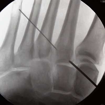
In the image above, a cannulated drill is being used to make a small hole through the cuneiform that will continue through the base of the second metatarsal. |
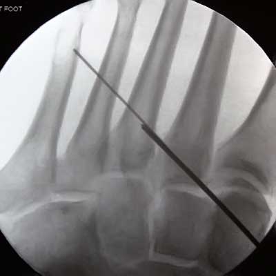
This x-ray shows the special suture button device which has been passed through the drill hole. The toggle button (two holes) is against the base of the metatarsal.. |
|
| |
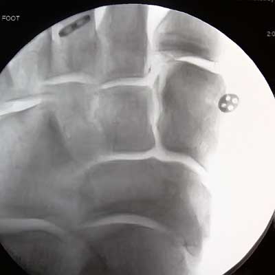
Now, the slide button (four holes) has been placed against the surface of the cuneiform bone on the right hand side. The Lisfranc interval is still gapping open. |
In the x-ray above, the surgeon is starting to apply tension to the suture that connects the 2 buttons. The gap between the cuneiform and the base of the second metatarsal is closing, and is approaching normal. |
|
| |
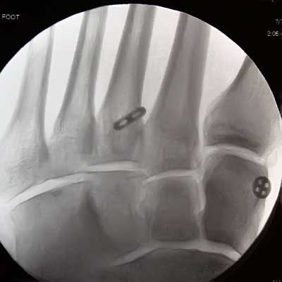
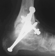
In the last image, the precise amount of tension has been applied to the suture connecting the buttons. As a result, the normal perfect alignment of the base of the second metatarsal and the cuneiform bone has been restored. The operation has been successfully completed. |
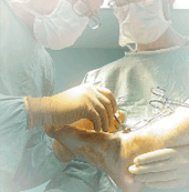
Post-operative Care:
The patient will not require a cast. The period of restricted weightbearing will be quite short. All patient's are carefully monitored to make sure that they have a successful outcome following surgery. This patient will have a much more rapid recovery due to the minimally invasive technique used.
Expert Care for You:
This complex and disabling Lisfranc injury requires expert care. Orthopedic foot and ankle surgeons have completed medical school and are Board Certified by the ABOS, and have additional highly specialized foot and ankle training. Orthopedic foot and ankle specialists have the most training in this area of diagnosis and surgery and are therefore well equipped to treat Lisfranc fractures as well as other complex foot and ankle conditions. Dr. DeGroot has extensive experience in nonsurgical, normal surgical, and minimally invasive surgical treatment for this injury. You may view his credentials here. If you would like more information on how these treatments may help you with your particular health concerns, please contact our office.
| Minimally invasive treatment is an alternative to fusion or more conventional fixation for ankle fractures, lisfranc fractures, heel bone fractures, metatarsal fractures, talus fractures, tarsal coalition, cavus foot, flat foot treatment, tibia fractures, some of which may be done with plates or screws. Minimally invasive surgery is best suited to a more active person. |
 |
|